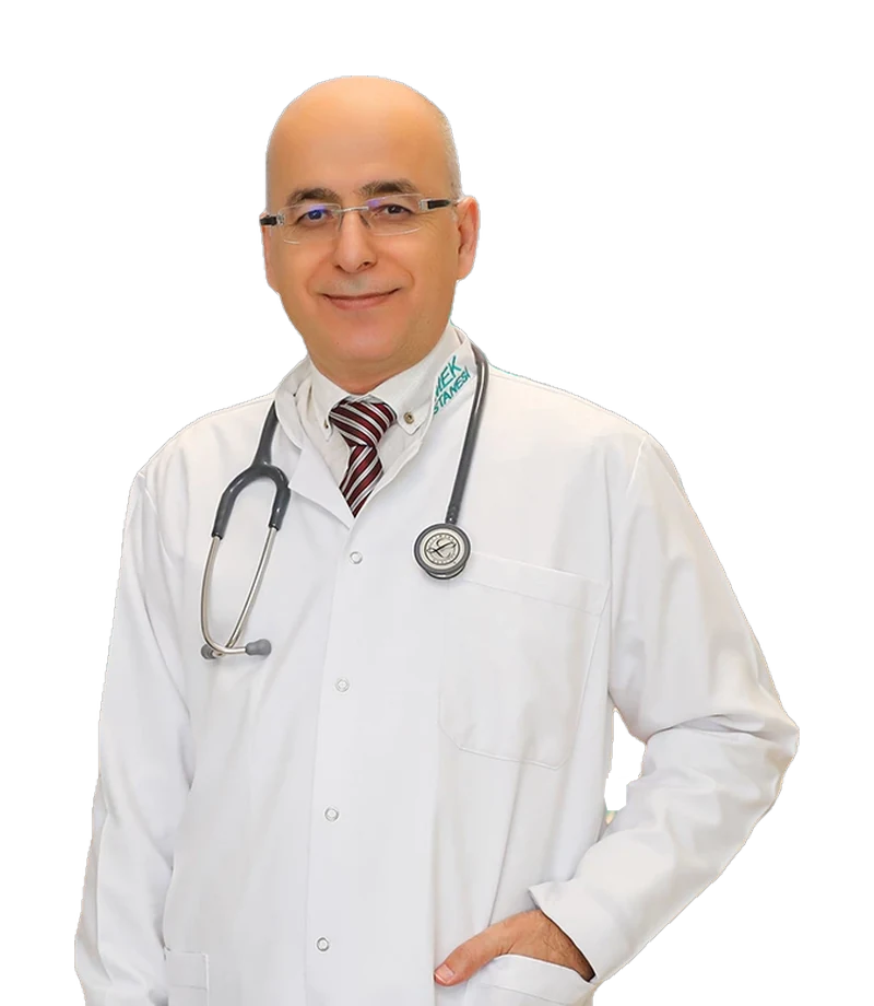
Spec. Dr.Hüseyin Biçer
Radiology
Book an appointmentAppointment
Not sure which department to go to?
Type your symptoms and we'll show you the right department and doctor.
Find a department by complaintDepartment
Radiology is a field that uses X-rays and other imaging methods for treatment purposes.
Our Doctors
Radiology
Book an appointmentSome of these imaging methods are ultrasound, computed tomography (CT), magnetic resonance imaging (MRI) and nuclear medicine methods. These imaging methods are used to determine how the disease will be treated. Radiology consists of two main branches: Diagnostic Radiology and Radiotherapy.
Radiology is also called the science of rays. The X-rays used in radiology can have harmful effects on a person if used excessively. For this reason, people working in this field wear protective clothing during imaging. X-ray films taken in radiology are very important in diagnosing many diseases. In some cases, a definitive diagnosis can even be made with X-ray films alone.
Radiology is the use of X-rays and other imaging methods in medicine for diagnosis and treatment. Some of the methods used for diagnosis and treatment include ultrasound, computed tomography (CT), magnetic resonance imaging (MRI), nuclear medicine methods, positron emission tomography (PET), mammography, fluoroscopy and certain other methods that use X-rays. When used for diagnosis, these methods make use of the images obtained to detect diseases; when used for treatment, they allow certain surgical procedures to be performed with less harm thanks to imaging. Radiology is divided into two main areas: “Diagnostic Radiology” and “Radiotherapy”. Some radiological methods are given below.
Ultrasonography: This method uses sound waves, not X-rays. It is used to image the internal organs of the abdomen (detecting stones, tumours and masses, examining the urinary and gynaecological systems, etc.). It has no side effects.
Doppler Ultrasonography: Used to image blood vessels.
Magnetic Resonance Imaging (MRI): When a specific magnetic field vector is dominant, the reflections of the energy sent with RF (radiofrequency) waves from the parts of the living body inside this magnetic field are converted into an image.
In general, this is the imaging of the body's blood vessels using contrast agents. It is used especially to image the blood vessels of the heart and brain.
Teleradiology is the digital transmission of radiology images between computers from one location to another via the internet or other connections. In this way, a radiology image taken very far away, even on another continent, can be evaluated and reported within seconds by a radiologist thousands of kilometres away. It is a very useful process for regions with a shortage of radiologists. In the years to come, PACS (picture archiving and communication system) will become very widely used.
Tomography was invented in 1915 by the French physician Boccage. Tomography is a medical imaging device that can produce images of sections of an organ between 1 mm and 10 mm thick. Tomography allows the body or organs to be examined from front to back, from top to bottom or in the horizontal sagittal plane. Tomography also involves radiation. Compared with radiological imaging, the patient is exposed to more X-rays and receives more radiation. Tomography usually brings Computed Tomography (CT) to mind. In fact, there is no difference between tomography and computed tomography; it is only a difference in name.
Thanks to advancing medical technology, tomography is constantly improving. The radiation dose given is being reduced and image quality is being improved.
Other departments